Turning Blurry Lines Into Brighter Views
What is Keratoconus?
Keratoconus occurs when the cornea progressively weakens, losing its round contour and forming a cone-shaped bulge. This irregular shape prevents light from focusing correctly on the retina, causing blurred or distorted vision. The condition typically starts in puberty and may persist into the mid-30s. Many patients are unaware they have keratoconus until their vision worsens.
Causes of Keratoconus
While the exact cause of keratoconus is unknown, several factors contribute:
- Weakness in the corneal structure
- Genetic predisposition (family history of keratoconus)
- Frequent or forceful eye rubbing
- An imbalance of corneal enzymes that makes the cornea more susceptible to damage
Symptoms of Keratoconus
The early stages of keratoconus may show subtle symptoms, such as:
- Slightly blurred or distorted vision
- Frequent changes in eyeglass prescriptions
- Poor vision not corrected with glasses
- Increased sensitivity to light and glare
- Double vision in one eye
Even when vision shifts, keratoconus doesn’t define your future, timely care does.
Even when vision shifts, keratoconus doesn’t define your future, timely care does.
Risk Factors of Keratoconus
You may be at higher risk if you have:
- A family history of keratoconus
- Chronic eye rubbing (often due to allergies)
- Connective tissue disorders such as Ehlers-Danlos or Marfan syndrome
- Conditions like asthma, hay fever, or eczema
- Poorly controlled allergies causing frequent eye irritation
How Keratoconus is Diagnosed?
Keratoconus is best detected with corneal topography, the most accurate diagnostic tool. This painless test creates a computerized map of the cornea’s surface, showing even subtle changes. Other tests, such as slit-lamp examination or corneal pachymetry (measuring corneal thickness), may also be used to confirm diagnosis and track progression.
Keratoconus Treatment
Treatment depends on the stage of the disease:
- Early stages: Corrective glasses or rigid contact lenses may improve vision.
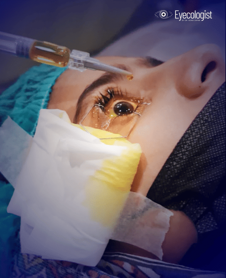
- Corneal Collagen Cross-Linking (CXL): A safe, one-time in-office procedure. A vitamin B (riboflavin) solution is applied to the eye and activated with UV light for 30 minutes, strengthening the cornea by forming new collagen bonds. Approved by the FDA in 2016, CXL is proven to stop or even reverse progression within 3–12 months.
- Advanced stages: If keratoconus progresses significantly, a corneal transplant may be required to restore sight.
Make an Appointment
Book Your Vision Journey Now